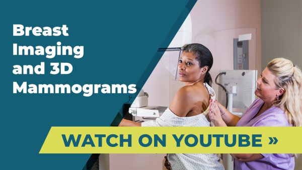
Mammography
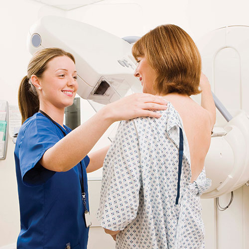
Mammography is an X-ray-based imaging test of the breast, considered to be the gold standard for breast cancer screening. It is designed to detect early-stage breast cancer in women experiencing no symptoms and to detect and diagnose breast disease in women experiencing symptoms such as a lump, pain or nipple discharge.
Screening and Diagnostic Mammography
There are two kinds of mammograms: screening and diagnostic. Screening mammograms involve two X-ray views of each breast. In the event an area of concern is identified, the patient may be called back for special additional views. This happens with about 7-10% of screening mammograms. The vast majority of these tests will be normal, although a small percentage of these follow-up (diagnostic) mammograms reveal breast cancer. For those in need, we offer a high-risk assessment program that provides information about available options.
In California, a doctor’s prescription is not required for you to have a screening mammogram. However, we are required to send a copy of the report to your primary care physician or OB/GYN.
Diagnostic mammograms require more views and are usually indicated in situations where patients have had prior breast surgery, implants or fibrocystic disease. In either exam, screening or diagnostic, the breasts will be temporarily and gently compressed in order to obtain the best possible image. To schedule your San Diego-area mammogram with Imaging Healthcare Specialists, please call us at (858) 658-6500.
3D Mammography (Tomosynthesis)
3D mammography, or tomosynthesis, is a recent and significant innovation in breast imaging—approved by the FDA in 2011. Numerous European and American clinical studies have demonstrated that adding tomosynthesis to a screening mammogram increases the cancer detection rate by about 40% and significantly lowers recall rates. It has also been shown to find more invasive cancers earlier than traditional mammography, which is why so many hospitals and imaging centers are adopting this technology. 3D mammography is also used for women with dense breasts. Dense breast tissue makes breast cancer screening more difficult and can increase the risk of breast cancer since dense breast tissue can mask potential cancer that may go undetected.


